However, the precise manner of the connection between the palatopharyngeus and the inferior constrictor has rarely been discussed, and its histological characteristics remain unclear. reported that the palatopharyngeus spreads radially and extends inferiorly. The evidence of these contributions has been anatomically explained based on its attachments to the soft palate, the pharyngeal raphe, the epiglottis, and the thyroid cartilage. The palatopharyngeus is one of the essential muscles required in proper swallowing, because it contributes to its various events, such as the movement of the soft palate, the shortening of the pharynx, and the elevation of the hyolaryngeal complex. The evaluation of mechanisms used by these anatomical structures may contribute to the treatment and rehabilitation of dysphagia.

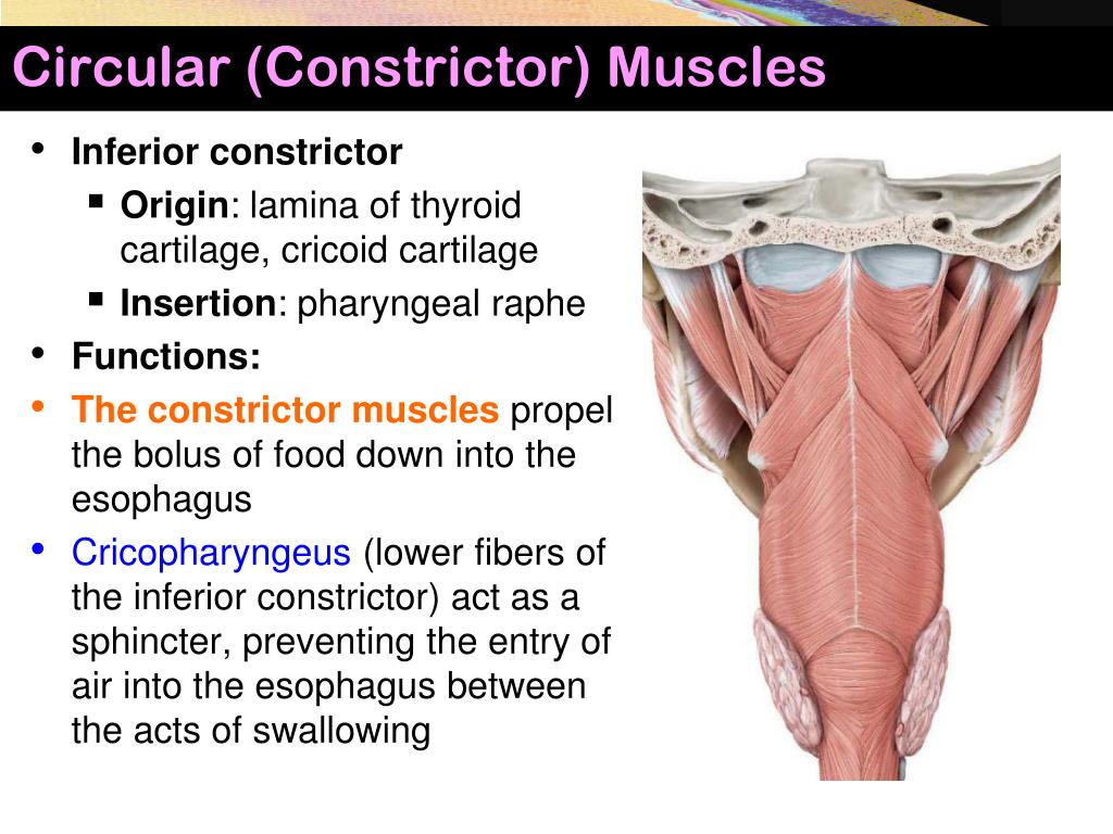
Therefore, the mechanism of the UES opening has been considered a passive event, and a few studies have focused on whether any anatomical structures directly act on the UES opening. In general, the UES opening occurs as a result of the elevation of the hyolaryngeal complex and the relaxation of the cricopharyngeal part of the inferior constrictor. The opening of the upper esophageal sphincter (UES), which is located on the cricopharyngeal part of the inferior constrictor and the uppermost part of the esophagus, is one of the essential events required for proper swallowing. The findings of this study show that the palatopharyngeus may act on the upper esophageal sphincter directly and help in its opening with the aid of the pulling forces in the superolateral direction. Therefore, the palatopharyngeus interlaced the cricopharyngeal part of the inferior constrictor through the dense connective tissues. The dense connective tissue not only covered the inner surface of the inferior constrictor but also entered its muscle bundles and enveloped them. Histological analysis showed that the inferior end of the palatopharyngeus continued into the dense connective tissue located at the level of the cricoid cartilage. The most inferior portion of the palatopharyngeus extended to the inner surface of the cricopharyngeal part of the inferior constrictor. Our observation suggests that the palatopharyngeus spreads radially on the inner aspect of the pharyngeal wall.

We examined 15 halves of nine heads from Japanese cadavers (average age: 76.1 years) 12 halves, macroscopically, and three halves, histologically. The purpose of this study was to examine the precise manner of the connection between the palatopharyngeus and inferior constrictor, and to examine the histological characteristics of this connection. Although previous reports suggest that the palatopharyngeus is partly interlaced with some parts of the inferior constrictor, the precise relationship remains unclear.

It is reported that the palatopharyngeus has muscle bundles in various directions and with attachment sites, and each muscle bundle has a specific function. The palatopharyngeus is one of the longitudinal pharyngeal muscles which contributes to swallowing.


 0 kommentar(er)
